
Cowbridge: 01446 773191
Cardiff: 029 2075 4087
Until relatively recently, the internal and external examination of eyes to detect signs of disease and deterioration was limited to shining a light onto or into the eye and examining the visible structures with a microscope. Whilst this is still and important skill for an eye care professional which can tell us about significant changes to our general and ocular health, it leaves a huge amount undiscovered, as the biggest clues to any changes lie beneath the surface.
OCT (Optical Coherence Tomography) is a highly sophisticated imaging technique, similar in principle to an MRI or Ultrasound scan.
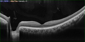
Our OCT scanner is able to generate highly detailed, three dimensional and cross sectional images of the structures within your eyes.
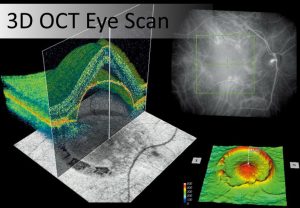
This advanced technique enables us to examine the vital parts or your eye in far greater detail than ever before, seeing beneath the surface of the retina to detect the earliest possible signs of sight threatening eye diseases such as glaucoma and macular degeneration.
By detecting these conditions years earlier than a conventional sight test would, we are able to initiate management and treatment sooner, which can greatly reduce the impact these conditions have on your sight.
An OCT scan is a quick and painless procedure taking only a few seconds. On some occasions we may need to put a drop in your eye to widen your pupil but we will discuss this with you if it is required.
We would recommend that all adults have an initial OCT scan so that we can record a baseline for your eye. We can then refer back to this initial exam in the future which makes detecting change easier.
As we get older our risk of developing sight threatening conditions such as glaucoma and macular degeneration increases significantly, more so if a close relative has suffered from one of these conditions.
For anyone aged over 40 with a family history of eye disease and for all those aged over 60 we would recommend that you have an OCT scan at every eye examination.
To return to examination options click here
